What is the liver?
...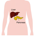
This is where the liver is situated
Functions of the liver?
...
How good your liver is functioning can be assessed by doing simple blood test called Liver function test (LFT). The component of which are:
Total Bilirubin
When total bilirubin is raised then it give yellow colour to your conjunctiva(eyes) and the condition is called Jaundice.
Liver enzymes (AST, ALT, ALP, GGT)
When ever there is any insult to the liver then alanine aminotransferase (ALT )and aspartase aminotransferase ( AST) are raised. But if there is any kind of blockage to the flow of the bile then alkaline phostatase (ALP)and gamma glutyl transferase (GGT)are raised.
Prothrombin Time
This test assess the clotting ability of the blood which is deranged in chronic liver disease commonly known s cirrhosis of the liver
Total Protein
Healthy liver produces albumin which is the main protein of the body. In In chronic disease, the liver’s ability to make proteins can be diminished, and this shows up by a fall in the serum albumin level.
Liver Cysts
Liver cyst is a common condition and usually detected incidentally while doing routine checkup. Nobody needs to be alarmed if this cyst is detected. You should consult your doctor for further evaluation.Mostly these are simple cysts which do not require any treatment. Simple liver cyst only requires treatment if they are symptomatic. There are no medications to decrease the size of the cysts, neither aspiration of the cyst is the treatment. These cysts can be easily tackled through laparoscopic approach in which a wall of the cyst is excised(Fenestration).
Occasionally, a liver cyst may have a solid component in the scan which signifies that it may be a cystadenoma or carcinoma . Then it requires urgent surgical consultation and further workup for excision.
Hydatid Cysts
Hydatid cyst of the liver is a common condition in India resulting from infection by a parasite known as Echinococcus. The usual causes of infection are eating uncooked/undercooked food, unwashed fruits/vegetables, and close contact with dogs. When humans pick up the infection, it results in formation of a parasitic cyst inside the liver and/or other organs including lungs, muscles, and brain.
People harbouring the cyst may be asymptomatic or present with dull abdominal pain. Cysts are normally seen on an imaging like ultrasonography, CT scan or MRI. They have a distinctive appearance on imaging but sometimes may require further investigations for confirmation of diagnosis. A blood test for antibodies to the parasite is frequently prescribed and and is indicative of infection. These cysts require treatment to prevent progression, spread and/or rupture. Treatment is primarily surgical.
During surgery either part of the cyst wall is removed (deroofing)after taking precautions to prevent any spillage or whole of the cyst ( pericystectomy) is removed. If there is any spillage of cyst fluid inside the abdomen, the infection may recur after sometime. We prefer laparoscopic pericystectomy in suitable cases or open deroofing. patients who are not candidates for surgery may be treated with injection therapy ( PAIR) or medicine ( albendazole).
Liver Abscesses
Liver abscesses are collection of pus inside the liver. It will present with pain abdomen, fever and fatigue. An ultrasound or CT scan can diagnose this problem. Treatment is antibiotics and sometimes ultrasound guided drainage of the pus. Rarely a liver tumour can present as an abscess.
Benign liver tumours
Haemangiomas
Hemangiomas are the most common tumours found in the liver. Nowadays many times they are found incidentally on ultrasound abdomen while being investigated for some other reasons. They are usually harmless and do not require any treatment; medical or surgical. Rarely they may become symptomatic and surgically treated with resection. Most of the times if you can prove convincingly that the tumour is a hemangioma you can safely forget it. A triple phase contrast CT scan or triple phase contrast MRI can confirm the diagnosis. Needle aspiration (FNAC) is usually not required.
Liver adenomas
These tumours ocuur primarily in females and associated with regular intake of Oral Contraceptive Pills (OCP). These tumours can turn into cancer in some patients. It is sometimes difficult to diagnose adenoma on the basis of imaging ( Ultasound/CT/MRI) alone. The treatment is surgical removal.
Focal nodular hyperplasia (FNH)
This is another type of tumour which can be diagnosed confidently with CT or MRI and can be left alone and observed. This type of tumour do not turn into cancers. Rarely in symptomatic cases surgical resection is indicated.
Primary liver cancer (HCC)
What is Hepatoma or Hepatocellular carcinoma (HCC)?
Hepato cellular carcinoma is a liver cancer which may develop in a healthy liver but more commonly in a cirrhotic liver. Patient developing cirrhosis due to hepatitis B/hepatitisC have more chance of having liver cancer and should have regular checkup with his doctor.
Liver cancer normally doesnot cause any sym0ptom when they are small only in advanced stage it can cause jaundice, weight loss, loss of appetite, low grade fever, lump palpable in the abdomen.
Diagnostic Test
Ultrasonography, Triphasic CT scan abdomen are the test to reach the diagnosis. Some time contrast MR and bone scan are required.
Alpha fetoprotien(AFP) is tumour marker.If the AFP level in the blood is raised, it very strongly suggests that the patient has a HCC. But the converse does not always apply – if the AFP levels are normal, that does not rule out a HCC.
Needle biopsy is not mandatory but can be done in few selected cases though it carries a small risk.
Treatment
It depends upon the the size of the tumour, location , number, it has spread to nodes, lungs /bones, whether liver is healthy or cirrhotic and fitness of the patient to undego operation if required.
If the liver is healthy, the patient fit, and the tumour can be safely cut out, then surgical resection of the tumour can be considered. If the tumour is large and cannot be safely removed, then chemoembolisation can be considered. If the tumour is small but the patient is unfit/unwilling to withstand surgery, radiofrequency ablation (RFA) can be considered. This involves placing a needle into the tumour and destroying it with energy generated at the tip of the needle.
If the liver is diseased (i.e. cirrhotic) but still in overall good condition, the patient is fit, and the tumour small, then liver transplantation should be considered. Surgical resection of the tumour or RFA may also be possible options. If the tumour is large and these options are not feasible, then chemoembolisation may be considered.
If the patient is unfit for surgery, the liver is very badly diseased, or the tumour is very large or has spread beyond the liver, then control of symptoms should be the main focus of care. Chemotherapy (in the form of intravenous injections or tablets taken by mouth) is not particularly effective in HCC, nor is radiotherapy.
Metastatic Liver Tumour
Metastatic Liver Tumour
Liver is a common site of secondary cancer i.e. Primary tumour is some where else but it has spread to liver. t is now well-recognised that surgical removal of the liver metastases from colon and rectum cancer can achieve a cure in a significant proportion of patients. When it comes to other cancers, surgical resection of liver metastases may still have a role to play, but only in a very small proportion of patients.
Another group of patients who may similarly benefit from liver resection are patients with liver metastases from neuro-endocrine tumours.
Diagnosis
Diagnosis is usually made on the basis of an ultrasound scan, and then a CT or an MR is done to confirm that. A whole body scan called FDG PET is useful in determining if there are other secondaries elsewhere in the body.
Treatment
Treatment of the liver secondaries depend upon the size of the tumour,condition of the liver, number and site of the secondaries and overall fitness of the patient.
Surgical resection of the tumour if indicated and can be done gives the best result. Other option are RFA, Chemotherapy.
Liver Resection
In a healthy liver upto two-thirds of the liver can be removed . Liver is an organ which can regenerates itself in six to eight weeks to its original size.Only if the liver is not healthy then we tend not to remove
a large portion of the liver.
All operation has got a risk. In major liver resection the risk of mortality /death is around 5% in other way chances of survival is 95%.
Having a liver resection
What is going to be done at the operation?
The most common operation we perform on the liver is to remove a part of it that has developed a tumour. This is called a liver resection (or a hepatectomy). The liver is made up of two halves called the right lobe and the left lobe. If the entire right lobe is to be removed, that is called a right hepatectomy, and if the left lobe is to be removed that is a left hepatectomy. Or the surgeon may just cut away the tumour, taking with it some surrounding normal liver tissue. If the tumour is near the gall bladder, or if the gall bladder is in the way, then the gall bladder is likely to be removed as well.
How does my body cope if I lose a part of my liver?
There is a lot of spare capacity in the liver. The liver can regenerate itself, and grows back to near-normal size within six to eight weeks. If your liver is healthy, you will be able to cope with removal of up to two-thirds of your liver. If there is cirrhosis or chronic liver disease, surgeons tend not to remove large portions of the liver.
What does having this operation involve?
Let us break that up into what happens before during and after the operation.
- Before the operation :
You will already have had different tests and scans. Once you are admitted for surgery, some more tests may be done, usually to confirm that you are well enough for the anaesthetic. These include blood tests, an ECG, sometimes a chest x-ray and tests of lung function. A member of the surgical team will have a discussion with you about the operation, after which you will be required to provide written consent for the operation to go ahead. - The operation itself :
A fairly large cut (or incision) will be made in your upper abdomen. The surgeon will expose the liver, assess that it is safe to remove the tumour, temporarily block the inflow of blood into your liver to reduce bleeding, and then cut away the part that is to be removed, using a special ultrasonic scalpel. Once the procedure is finished and all bleeding has been controlled, blood flow to the liver will be restored and the wound stitched up. - After the operation :
When you wake up, you may find yourself in the intensive care or high dependency unit, or you may be back in the ward. There will be an oxygen mask on your face. In addition to the drips going into your forearms and your neck, the tube in your nose and the urinary catheter, there will be one or two plastic tubes (drains) emerging from your tummy, which will remove any unwanted ooze You will probably feel some pain and sickness, but we will give you medicine for this.
Can something go wrong?
Every operation carries some risks, and so will yours. A general anaesthetic carries some risk, and there is a small chance that you may develop problems relating to your heart or your breathing. If you already have heart disease or lung disease, this risk is increased. The various lines (needles) and catheters that are put in may cause some bleeding or local injury or may introduce infection. They are, however, put in with great care and with sterile precautions.
Bleeding during or after the operation may happen after any surgery. Blood will be cross-matched and kept available should you need a transfusion. Transfusion itself carries some risks (reactions from a mis-match, transmission of viruses) and is not given except when absolutely necessary. Most operations on the liver do not require a blood transfusion.
Infection is another possible complication, and can involve the wound, or the inner organs. You will receive antibiotics around the time of surgery to try and prevent this from happening. The risk is increased if any bile leaks from the cut surface and collects deep to the wound.
If you have a large portion of your liver removed, sometimes you may develop a degree of liver failure in the immediate aftermath of the operation. This may make you jaundiced for a time, and interfere with your blood clotting. Such liver failure is usually short-lived and the liver makes up for the loss in a matter of days or weeks.
The pain from your wound will make coughing difficult. Patients sometimes develop chest infection from retained phlegm. Deep breathing, clearing out the phlegm in your throat and chest and working with the physiotherapist is very important after your operation. Lying still in bed can lead to the formation of clots in the legs (called deep vein thrombosis or DVT) and these can sometimes float off in the circulation and reach the lungs (called pulmonary embolism or PE). These are serious complications, and we try hard to prevent them. You will be asked to wear elastic stockings, and will receive a mild dose of an anti-coagulant to reduce the risk of clot formation. You can help by moving your legs in bed, and getting out of bed as soon as your condition will allow after the operation.
What are my chances of surviving the operation?
Information collected from hospitals all over the world indicates a mortality rate of less than 5% from major liver surgery. In other words, you have at least a 95% chance of surviving your operation.
Will I be cured?
If the operation is to remove a cancer, that question is difficult to answer immediately. After the operation we would have to wait and see. Liver resections for cancer are usually carried out with the intent to cure. But there will undoubtedly be a significant risk of the tumour coming back, and only time will tell if you have been cured. The pathologist’s report on the pieces of tissue that are removed will give us some clues.
Sometimes, the surgeon may find that the cancer cannot be safely removed. For example, the growth may be larger than the scans suggest, or there may be some other obstacle in the way. In such situations, the aim will be to do everything possible to relieve your current symptoms and prevent future problems. This may involve the creation of a join between the bile duct and the bowel to relieve or prevent jaundice (and this goes with an additional join between bowel and bowel). This is often referred to as a biliary bypass procedure.
Your operation may be carried out not for cancer but to relieve symptoms caused by a benign tumour. Jaundice is usually relieved quite successfully. In the case of chronic pain, it is difficult to predict how successful the operation will be in relieving pain. You would have to wait and see.
Portal Vein Embolisation (PVE)
What is PVE?
Sometimes, the tumours are located in such a manner within the liver that a large part of the liver needs to be removed. For example the entire right lobe and part of the left lobe may need to be removed – which is called an extended right hepatectomy. But the amount of liver that will then be left behind may be too small and the patient will run a very high risk of developing liver failure. This applies particularly to situations where less than 25% of the total volume of the liver is likely to be left behind (or, in patients with chronic liver disease, less than 40% of the liver will be left behind). In such situations, it is possible to block off the portal vein inflow of blood into the parts of the liver that are going to be removed. They then start to shrink (atrophy) while the rest of the liver (the part that is going to be left behind) starts to grow. In a period of 2 to 6 weeks, substantial growth may be seen, and a surgical resection may become possible.
How is it done?
The procedure itself involves a puncture of the liver to inject glue-like material into the relevant branch of the portal vein. This done in the x-ray department by a radiologist, using ultrasound and angiography techniques to direct the needle into the correct position Alternatively the branch of the portal vein may be tied off during a surgical operation.
What are the risks?
There are some risks involved, including bleeding or bile leak from the puncture into the liver. Spillage of the glue into parts of the circulation is another small risk. It is difficult to predict exactly how much liver growth this procedure will cause in a particular patient, and one has to wait and see.